8 Arms and Legs
Objectives
- Identify the fin and limb elements of the major vertebrate clades.
- Orient the limb elements.
- Study the fusion and reduction of distal limb elements in relation to locomotor adaptation.
Overview
In this lab you will continue the exploration of the appendicular skeleton. The limbs of vertebrates perform functions in locomotion, support, maneuverability, stability, and object manipulation. The selective pressures from these activities shape the limbs, resulting in a large amount of morphological variation in vertebrates. Not all of the listed features can be found in all animals. Most of the structures listed below are most readily found in mammals, and that is a good place to start.
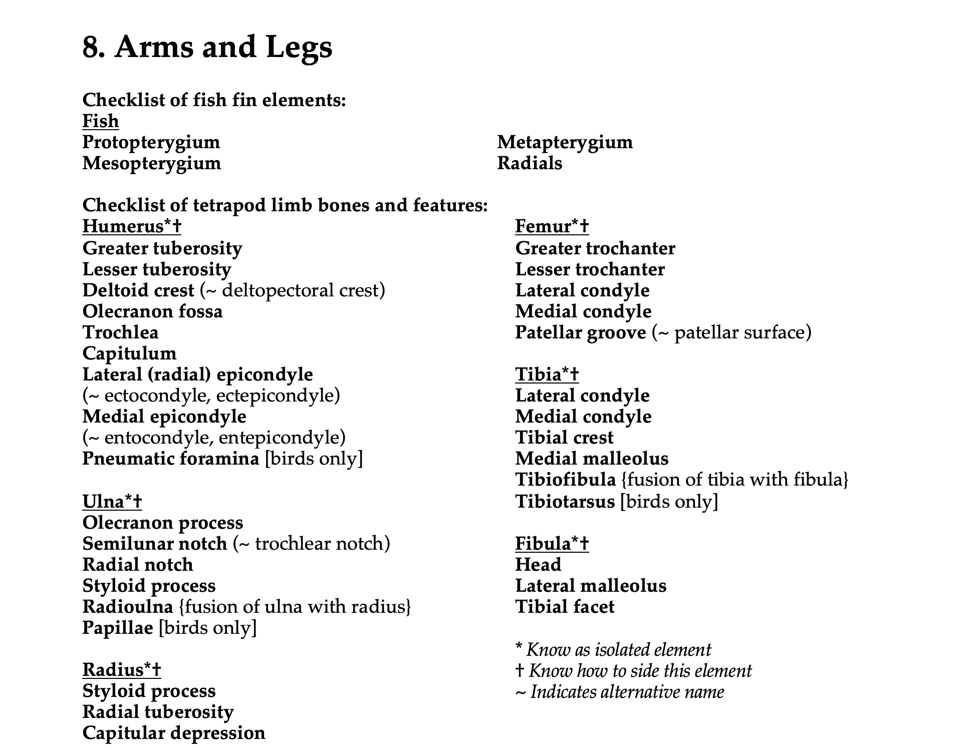
See the terms list here: Terms 8.1
From fins to limbs
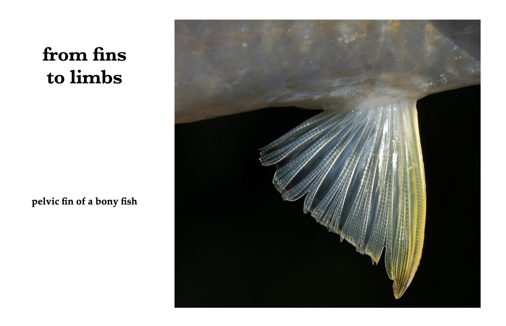
Figure 8.1. Pelvic fin photo from Wikipedia user W.A. Djatmiko under CC BY-SA 3.0.
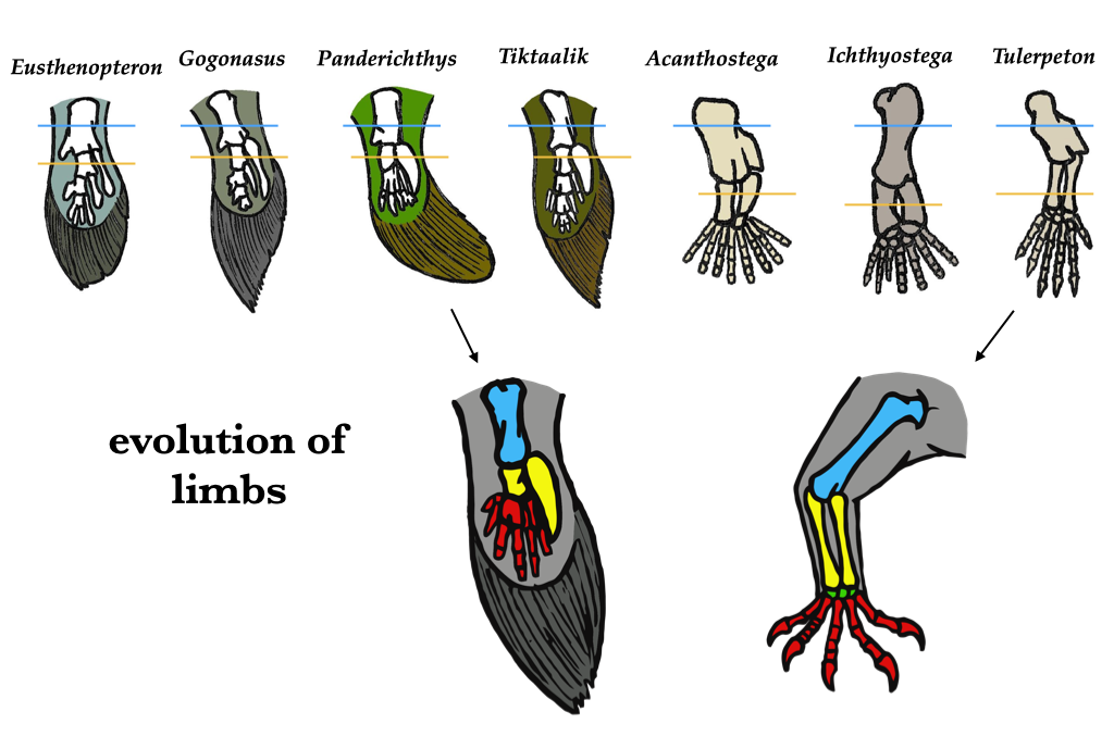
Figure 8.2. Comparison between fins and legs from Wikipedia user Conty under CC0 public domain.
Chondrichthyes
Living sharks have three basal cartilaginous pterygiophores (the basals) that articulate with the pectoral girdle in the glenoid region: an anterolateral propterygium, an intermediate mesopterygium, and a posteromedial metapterygium. Segmented radial pterygiophores (radials) extend from the basals, and fin rays extend from the radials. The pelvic fin typically possesses an anterior propterygium and a posteromedial metapterygium, with radials and fin rays distal to those.
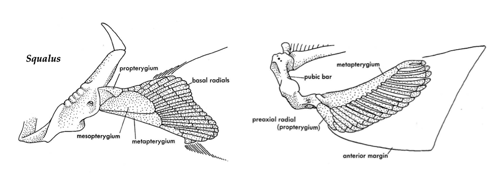
Figure 8.3. Pectoral (left) and pelvic (right) fin bases of a shark (Squalus). Illustrations from Jollie (1962) under CC0 public domain.
Actinopterygii
Actinopterygians have three small basal pterygiophores with many radials. The radials provide the majority of the support for the limb. The radial on the anterior (dorsal) fin margin is the largest, and it is usually cartilaginous, resembling a propterygium. In amiids and sturgeon, the radials are associated with a metapterygium.
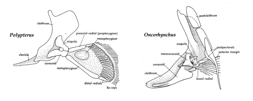
Figure 8.4. Pectoral girdles and fin bases of a bichir (Polypterus) and a salmon (Oncorhynchus). Illustrations from Jollie (1962) under CC0 public domain.
Lissamphibia
Caudata
Salamanders have a humerus, radius, and ulna in the forelimb, and a femur, tibia, and fibula in the hindlimb. The distal pair of limb elements are approximately equal in size.
Anura
The limbs of frogs are specialized. The radius and ulna fuse to form a radioulna, and the tibia and fibula fuse to form a tibiofibula. Humeri of male anurans are frequently modified by several large crests to which large muscles attach. The anuran hindlimb is long relative to the forelimb, and it is bolstered by a fused astragalus (tibiale) and calcaneum (fibulare). The jumping ability of frogs is based on the elongation and strength of these limb elements.
Testudines
Turtle humeri vary widely in form, a not surprising fact given the extent of turtle locomotor diversity. The shaft of the turtle humerus is typically held horizontally, and the humeral head is oriented (almost) vertically, and it has prominent tuberosities on both sides. On the sides of the femoral head, the shaft forms large trochanters and, distally, the shaft expands into fibular and tibial condyles. The tibia is larger than the fibula in turtles.
Crocodylia
The crocodile humerus has a relatively large deltopectoral crest. The ulna does not have a distinct fossa at the elbow.
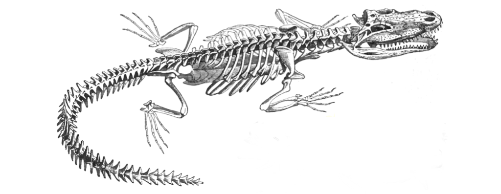
Figure 8.5. Crocodile skeleton from Lydekker (1896) under CC0 public domain.
Aves
Bird limb bone morphology shows less variation than other taxa because most birds use flight as the primary means of locomotion. From the side, the shaft of a bird humerus forms an attenuated S. The proximal end is larger than the distal end, with a large deltoid crest present just distal to the lateral border of the humeral head. One or more pneumatic foramina occur on the posterior surface. The distal end of the shaft is concave anteriorly and convex posteriorly, and the lateral external condyle is the larger. To side a bird humerus, follow the advice of Gilbert et al. (1981, p. 70): “Hold the bone vertically, proximal end up, pneumatic foramen facing the observer, bicipital surface facing away. The deltoid ridge is on the side the bone is from.”
The proximal end of the radius is concave; the distal end is flared and broader than the shaft. To side a radius: “Hold the radius so that the proximal end is toward the observer, the radial tuberosity faces down, away from the observer. The radial shaft is bowed so that it is concave on its medial edge, convex laterally. The styloid process is nearest the mesial side.” (Gilbert et al. 1981, p. 100).
From a lateral view, the avian ulnar shaft is ventrally concave. The dorsal surface has papillae for feather attachment. The proximal end has a large medial facet and a smaller lateral facet. The distal end has a styloid process. To side an ulna: “Hold the bone with the proximal end toward the observer, the semi-lunar notch (ventral surface) facing up. The smaller facet is lateral, on the side the bone is from. The styloid process points medially, away from the side the bone is from.” (Gilbert et al. 1981, p. 116).
Bird femora have a convex anterior surface and a large greater trochanter that is as high or higher than the femoral head. To side a femur: “Hold the bone vertically so that the head is up, the patellar groove facing away from the observer. The greater trochanter and the epicondyle are both lateral, on the side the bone is from.” (Gilbert et al. 1981, p. 160).
The tibia, astragalus, calcaneus, and sometimes the patella are fused to form the tibiotarsus. To side a tibiotarsus: “Lay the bone on a flat surface with the proximal end toward the observer, the anterior surface facing up. The fibular crest and outer cnemial crest are lateral, on the same side the bone is from. The shaft curves medially to meet the medial condyle on the opposite side the bone is from. The medial condyle is almost always larger in anterior-posterior diameter than is the lateral.” (from Gilbert et al. 1990, p. 184).
Mammalia
Unlike birds, mammal limb morphology is highly variable. The long bones of mammals have epiphyses that generally fuse completely to the diaphysis in adult placentals, but they remain unfused in juveniles and some marsupials.
The head of the humerus is rounded and is easily distinguished from the trochleated distal end. There is often an entepicondylar foramen.
To side a humerus, “When in anatomical position, the head of the humerus is proximal and nearest the midline of the animal, where it articulates with the scapula. An intertubercular groove for the tendon of the biceps leads down from and anterior to the head. It is on the medial side of the shaft. Viewed from the front, if the groove is to the observer’s right then the bone is from the animal’s right side, and vice versa.” (Gilbert et al. 1981, p. 44).
The radius and ulna together allow a great range of motion, as the radius is able to rotate across the ulna in supination. In some groups, such as ungulates and lagomorphs, the radius and ulna are fused, preventing rotation.
The head of the femur is much more rounded than that of the humerus and the distal end is trochleated.
The mammalian tibia is a large bone, and the fibula is often reduced or absent. The tibia and fibula can be distinct or can have a variable amount of fusion along their length, depending on lineage.
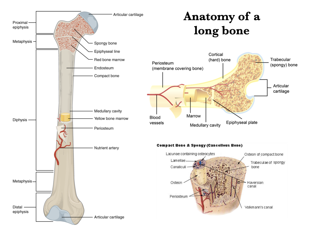
Figure 8.6. Anatomy of a long bone from Wikipedia user OpenStax College under CC BY 3.0; Bone cross-section from Wikipedia user Pbroks13 under CC BY 3.0; Compact and spongy bone from Wikipedia user SEER under CC0 public domain.