10 Functional Morphology
Objectives
- Understand how fore- and hindlimbs function during tetrapod locomotion.
- Gain experience in measuring morphological parameters.
- Identify and describe both similarities and differences in morphology among animals that use differing locomotor modes.
Overview
In this lab, you are going to explore the relationship between locomotor mode and morphological variation. The bones and muscles of limbs together create lever systems that are used to propel the animal through its environment. The properties of these levers reflect the environment in which the animal must maneuver because different environments impose different demands on the animals that use them. To explore these properties, you will describe the positions and characteristics of muscle attachments in a variety of diverse animals and subsequently relate them to the locomotor modes that these animals perform. You will be taking measurements for 8 muscles (described below) on each of a minimum of 10 species that represent different locomotor modes (listed below).
Materials
A measuring device. We will provide rulers, string, etc. which can be found at the front of the lab. You will find that some devices are more effective than others for a given animal.
A datasheet. You will be recording a lot of information and it will be to your benefit to have it well-organized.
Locomotor modes
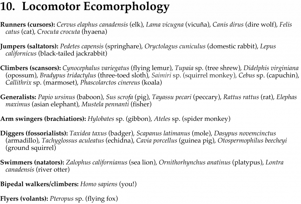
Below is a list of species sorted by locomotor mode. From this list, select at least 10 species. You can choose whichever species you would like however you must include 3+ species from at least 3 of the locomotor categories (so that you can compare within as well as among locomotor modes). The 1 remaining species can be from any locomotor mode.
See the list here: Terms 10.1
Measuring in- and out-levers
The muscles that you will be measuring are listed below. For each muscle, identify the lever type (1, 2, or 3) and measure the lengths of both the in- and out-levers in all 10 species you selected from the list above. As you are doing this, think carefully about which parts of the limb represent the different components of the lever. For example, when measuring the out-lever, you should account for the action of the muscle, that is, what joint it is moving, and where the force is likely to be exerted to resist that motion. Since these muscles all pertain to locomotion, it is reasonable to assume that the out-force will be located on the foot, but the specific location on the foot will depend on foot posture and how the foot is to be used. Be sure to take note of WHERE and WHY you used the locations for in-force, out-force, and fulcrum that you did. We strongly encourage you to make sketches (even if crude) of this for your own reference and benefit.
Remember that the length of the out-lever is the distance between the fulcrum and the location of the out-force, and the length of the in-lever is the distance between the fulcrum and the location of the in-force.
biceps brachii
Origin: edge of the glenoid fossa
Insertion: bicipital or radial tuberosity (on the radius)
Action: flexion of the arm at the elbow joint
triceps brachii
Origin: axillary border of the scapula (long head), deltoid ridge of humerus (lateral head), posteromedial side of the proximal end of the humerus (medial head)
Insertion: proximal end of the olecranon process
Action: extension of the arm at the elbow joint
teres major
Origin: caudal edge of the scapula
Insertion: medial side of the humerus (directly opposite of the deltopectoral ridge)
Action: adduction of the humerus (moves humerus medially)
semimembranosus
Origin: ischial tuberosity
Insertion: medial epicondyle of the femur and proximal end of the medial surface of the tibia
Action: extension of the thigh at the hip joint (moves the femur caudally)
rectus femoris
Origin: ilium
Insertion: patella, and thereby into the tibial tuberosity
Action: flexion of the thigh at the hip joint (moves femur cranially)
vastus lateralis
Origin: greater trochanter
Insertion: tibial tuberosity
Action: extension of the lower leg at the knee (moves the lower leg anteriorly)
biceps femoris
Origin: ventral surface of the ischial tuberosity
Insertion: proximal end of the posterolateral surface of the tibia
Action: flexion of the lower leg at the knee (moves the lower leg posteriorly)
gastrocnemius
Origin: lateral condyle and medial condyle of the femur
Insertion: proximal edge of the calcaneum
Action: extends the foot at the ankle
In- and out-levers affect limb function
The relative sizes of the in- and out- lever arms affect both force (Forcein x Leverin = Forceout x Leverout) and velocity (Velocityout x Leverin = Velocityin x Leverout) of the limb. Think about the locomotor modes that you measured in terms of out-force production and of out-velocities relative to each other. Other variables that might help in thinking about and discussing these modes are mechanical advantage and gear ratio. Mechanical advantage is an increase in out-force associated with an increase of in-lever length (Forceout = (Forcein x Lengthin)/Lengthout). Gear ratio (GR = Lengthout / Lengthin) is a relationship between force and velocity, where higher values indicate higher velocities. Calculate gear ratio for each species that you measured. Rank them from lowest gear ratio to highest.
Describing evidence of muscle size and force
The size of a muscles is often indicative of the force that it can produce, but there are many times in which muscles are no longer present with skeletal specimens. We can examine the “scar” that the muscle left on the bones where it was attached. Larger, more distinct scars that are rough in texture and appear to be composed of particularly thick bone typically mean larger and more powerful muscles were once attached there. On the contrary, smaller muscles leave scars that have a smaller area, smooth texture, and bone that is not noticeably thicker than that surrounding it. We do not expect you to measure muscle scars in this lab, but this information may help you make the length measurements that are expected of you and could provide inspiration for your projects.
The Writeup
You will each turn in an individual writeup of this lab. Even though you worked with a partner to collect the data, you will complete the writeup by yourself. This should have an overall format that mirrors a scientific paper. We don’t expect you to read a ton of literature for this, but your lecture notes and reading materials will likely be good resources for background information. If you are not confident with this kind of science writing, come talk to one of us in office hours, or if you have extra time at the end of lab. Do NOT wait and email the night before the writeup is due; that would be too late.
You should begin by explaining what the main concept is you’re exploring, why it’s interesting, and lead into a hypothesis about this concept (introduction). You should then write about what you did and why you did it that way (methods). Then describe the data you collected but wait until the discussion section to talk about what it means (results). Last, talk about what the data means and what biological processes are involved with your results (discussion). The focus should be on the results and discussion; those are the interesting bits.
In your writeup, make sure you address the following questions:
- What kind of lever system is involved for each of the muscles you’re studying here (type 1, 2, or 3)? For what reason do you think that type evolved here, and not another one?
- For the species with the highest gear ratio, the lowest gear ratio, and one somewhere in between, make predictions about their natural history and why having such gear ratio might be advantageous.
- What are the differences between the lever systems for different muscles? Between the arms and legs? Between different locomotor types? Use lever mechanics to explain why the locomotor types you looked at differ the way they do. Describe why these differences in force and velocity production might be evolutionarily advantageous.
