2 Fish Skulls
Objectives
- Gain familiarity with the basic regions of the vertebrate skull—neurocranium, splanchnocranium, dermatocranium.
- Identify the basic components of the shark chondrocranium.
- Identify the elements of the fish head listed below and recognize the skull region to which each element belongs.
- Recognize which bones are endochondral and which are intramembranous in origin.
Overview
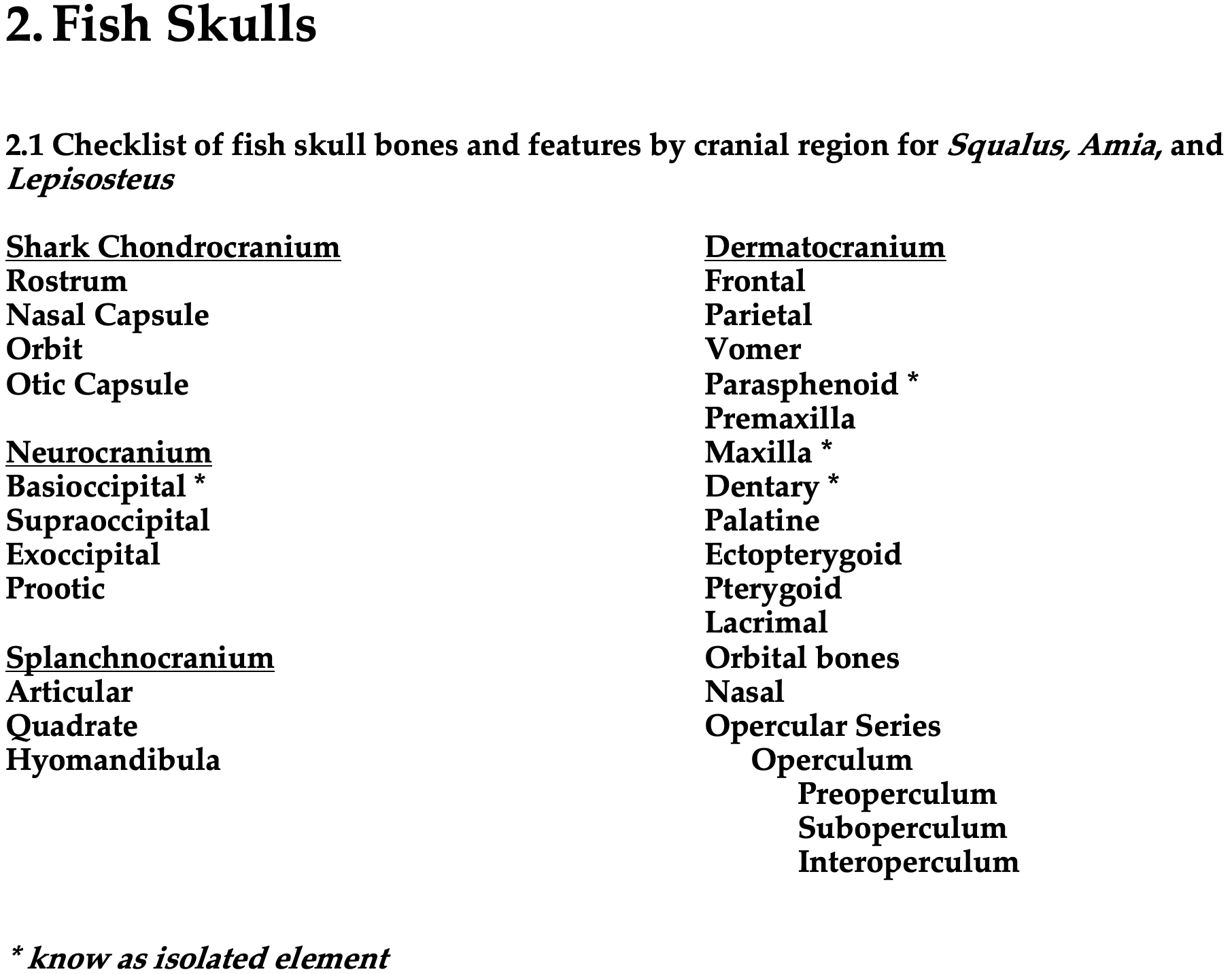
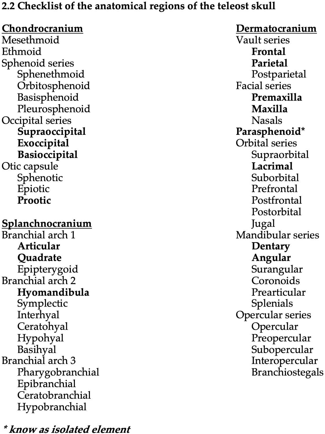
This lab marks the start of a study of the vertebrate skull. Vertebrates are unique in having a distinct head where the sense organs, brain, mouth, and gills are located. We will spend a few labs on the skull, beginning today with the “fishes,” including the most basal living forms (lamprey and hagfish); the cartilaginous forms such as sharks and rays (Chondrichthyes); and bony fish such as carp (Actinopterygii). After this lab, you will be able to identify the bones and features of the fish skull listed below (Table 2.1) in bold face. You only need to know these objects in the context of the articulated skull, except for those with an asterisk (*), in which case you should try to learn to identify the object in isolation as well.
See the terms list here: Terms 2.1–2.2
Strategies for success
You will learn the skull bones of many different taxa, and it is important to compare the skulls of fishes from different groups to understand the relative shapes and positions of the different elements. The study of morphology is always comparative, and you can take advantage of the diversity of specimens available in lab as well as the figures herein. Homologous elements have conserved shapes and articulation patterns, and the study of bone shape and relative position across taxa can be your main focus.
The vertebrate cranium has three fundamental parts
Chondrocranium
The cartilaginous chondrocranium is the underlying scaffold of the skull, and it forms the entire skull in the chondrichthyans. In the osteichthyans, the cartilage of the chondrocranium is ossified (replaced by endochondral bone), and it is referred to as the neurocranium. In teleosts and tetrapods, the neurocranium forms the central base of the skull.
Splanchnocranium
The splanchnocranium is formed by the branchial arches (gill arches). In some vertebrates, the branchial arches make up part of the hyoid apparatus and jaw mechanism. The splanchnocranium is made of endochondral bones.
Dermatocranium
The dermatocranium is composed of the dermal bones that form the superficial parts of the skull.
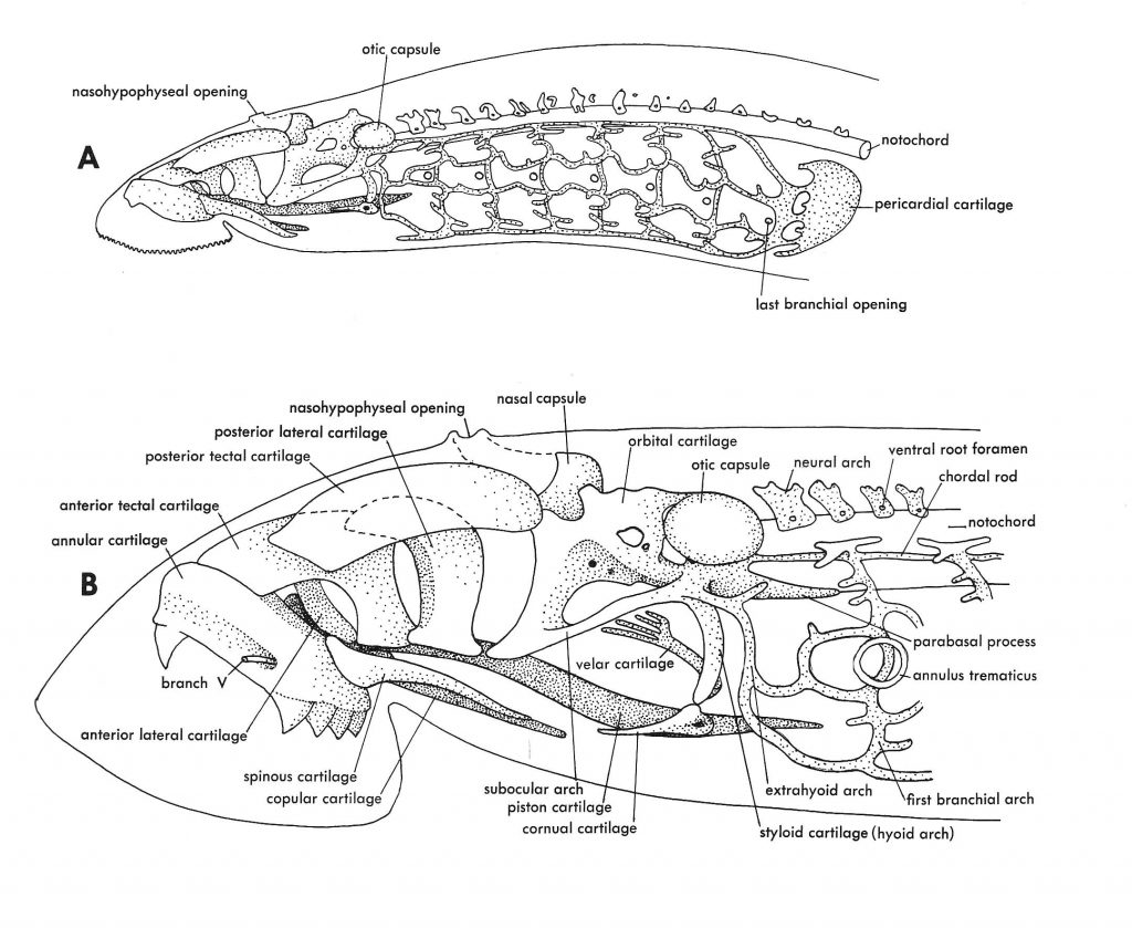
Figure 2.1. Cranium and visceral skeleton of a lamprey as an example of a primitive vertebrate skull. From Jollie (1962) under CC0 public domain.
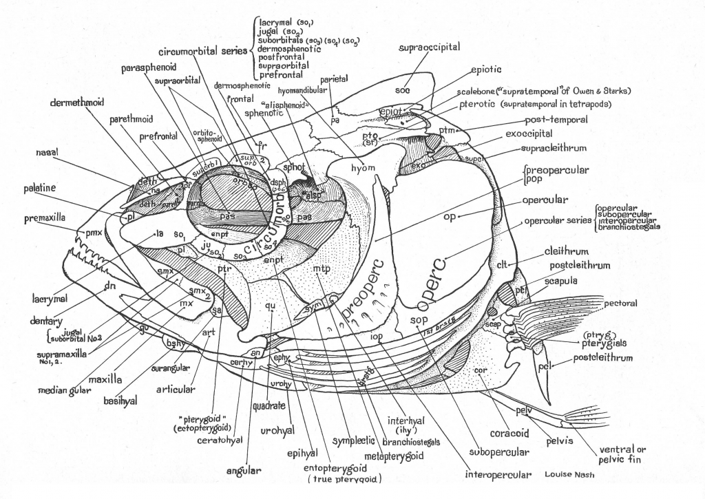
Figure 2.2. Idealized bony fish skull from Gregory 1933 under CC0 public domain.
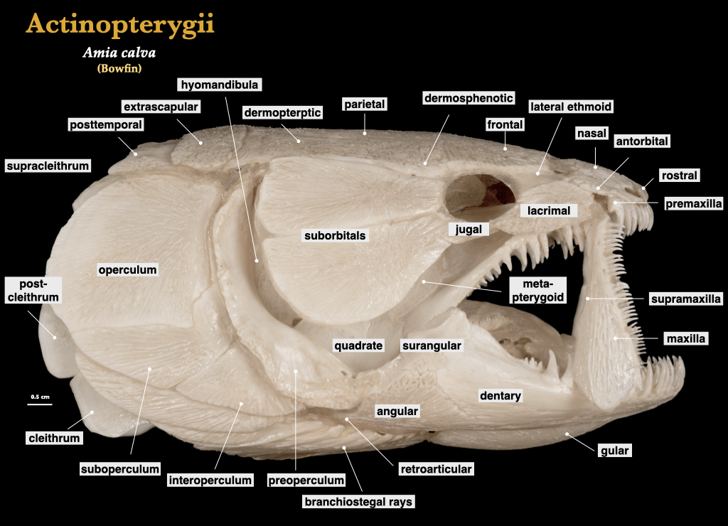
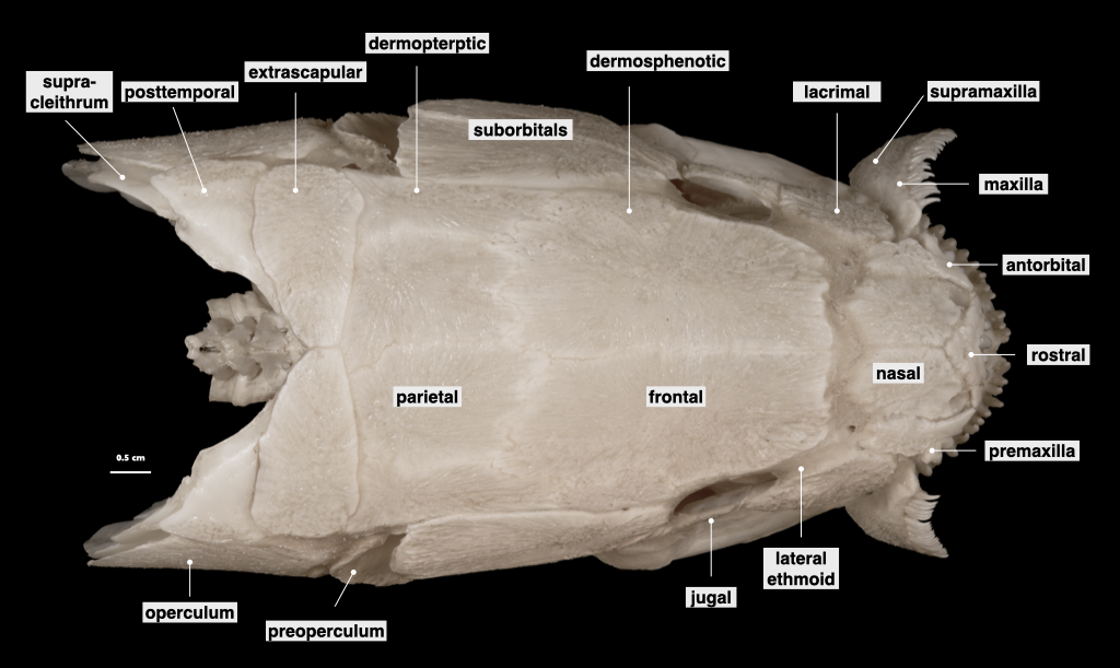
Figure 2.3. Skull of the bowfin (Amia) in lateral and superior views. Amia calva, private collection.

Figure 2.4. Illustration of bowfin (Amia) skull by Lauren C. Sallan, used with permission; bowfin photo from Ellen Edmonson and Hugh Chrisp under CC0 public domain.
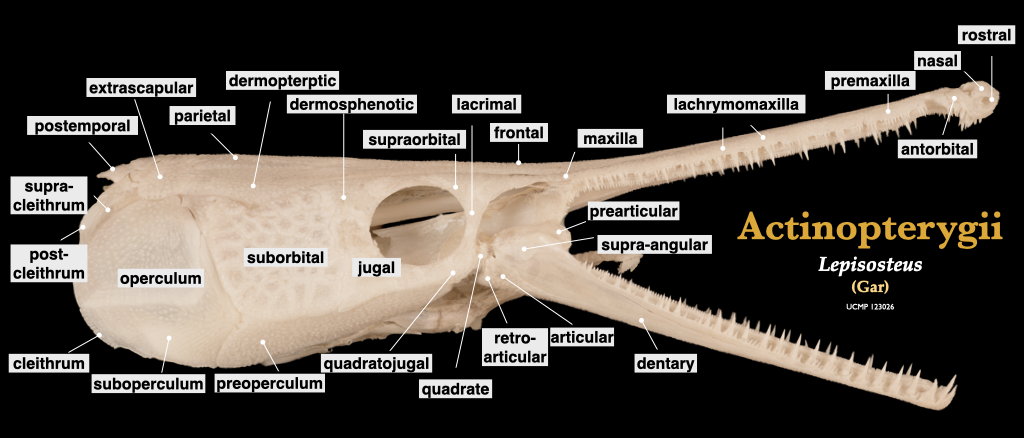
Figure 2.5. Skull of a gar (Lepisosteus) in lateral view. Lepisosteus specimen, UCMP 123026.Figure 2.6. Illustration of gar (Lepisosteus) skull by Lauren C. Sallan, used with permission; gar specimen photo from Wikipedia user PierreSelim under CC BY-SA 3.0.
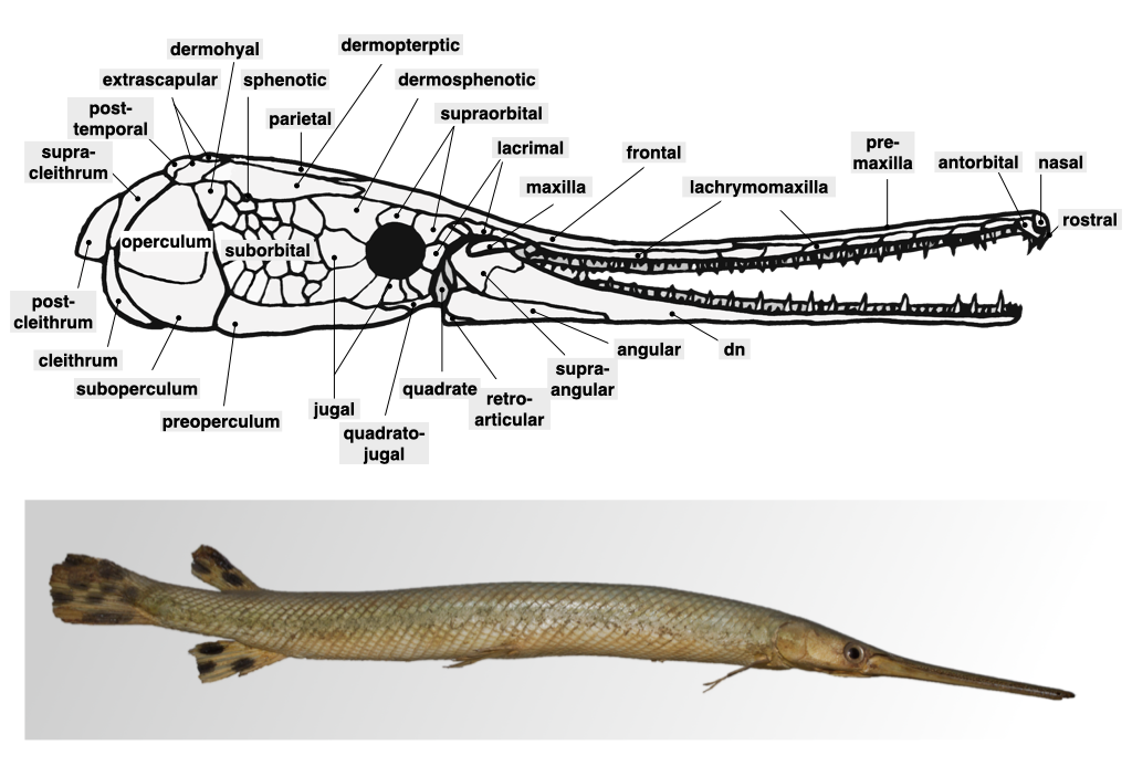
Figure 2.6. Illustration of gar (Lepisosteus) skull by Lauren C. Sallan, used with permission; gar specimen photo from Wikipedia user PierreSelim under CC BY-SA 3.0.
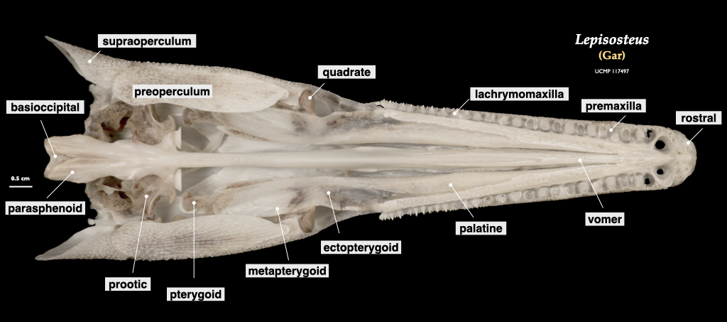
Figure 2.7. Cranium of a gar (Lepisosteus), inferior view. Lepisosteus oculatus, UCMP 117497.
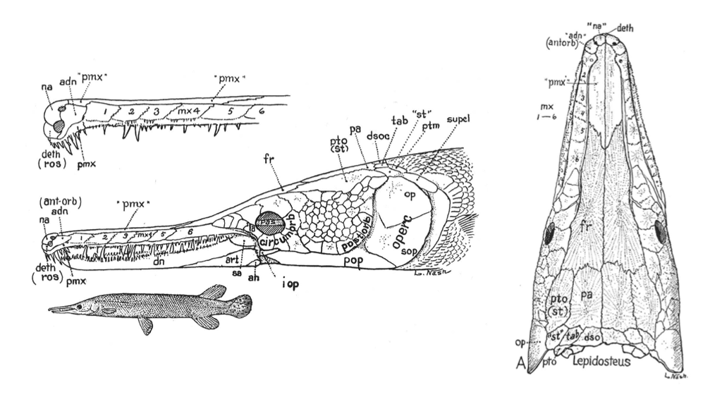
Figure 2.8. Skull bones of a gar (Lepisosteus) from Gregory (1933) under CC0 public domain.

Figure 2.9. Skull of a carp (Cyprinus carpio), lateral view.
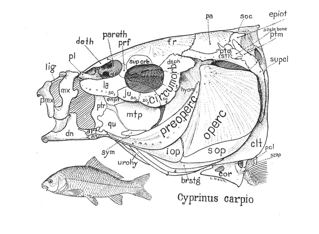
Figure 2.10. Skull bones of a carp (Cyprinus carpio) from Gregory (1933) under CC0 public domain.