9 Wrists, Ankles, Hands, and Feet
Objectives
- Identify the major bones of the tetrapod manus and pes both in context and disarticulated.
- Orient the carpometacarpus and tarsometatarsus of birds, the tarsus of crocodilians, and the major mammal elements of the carpus and tarsus.
- Recognize the standard autopodial postures.
- Understand and be able to write and interpret phalangeal formulas.
Overview
Wrists, ankles, hands, and feet are all part of the tetrapod autopodium. Throughout evolution, the autopodium has undergone extensive modification. Each autopodium has some number of rays. Each ray contains a single metapodial and a series of phalanges that extend distally. The rays attach proximally to several distinct bones, the podials. The number of rays in the autopodium, and the morphology of the autopodium as a whole, correlate with the functional demands imposed by lifestyle. In this lab, you will learn the morphology of the vertebrate autopod and explore autopod diversity across taxonomic groups and locomotor modes. By the end of this lab, you should be able to identify all elements in articulation and the major elements in isolation.
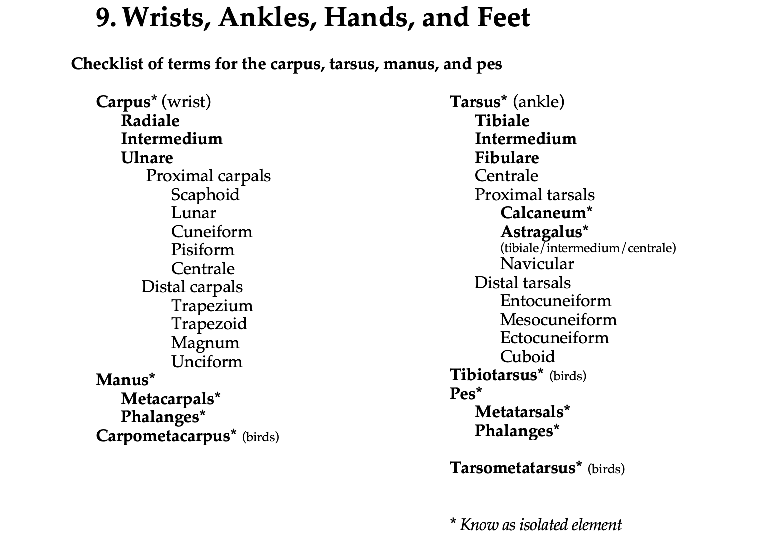
See the terms list here: Terms 10.1
Hints and guidelines
A couple of terminological points: the manus includes the metacarpus and the phalanges of the hand, and the pes includes the metatarsus and the phalanges of the foot. The carpus refers to the bones in the wrist and can be subdivided into proximal and distal carpal groups. The tarsus refers to the bones of the ankle and may also be subdivided into two groups. Podial is a general term that refers to both carpal and tarsal elements, and metapodial is a general term for both metacarpals and metatarsals. A ray includes the metapodial plus the phalangeal series. A digit includes only the phalanges.
Nomenclature of carpals and tarsals
| General terms | Mammalogy | Human Anatomy |
| Carpals | ||
|
|
|
|
|
|
|
|
|
|
|
|
|
|
|
|
|
|
|
|
|
|
|
|
|
|
|
| Tarsals | ||
|
|
|
|
|
|
|
|
|
|
|
|
|
|
|
|
|
|
|
|
|
|
|
|
1 Also called Os multangulum majus, 2 Also called Os multangulum minus
Evolution of the digits and a numbering scheme
Each digit is identified by a number, typically represented by a roman numeral. By convention, digits are numbered from medial to lateral on each limb. Thus, in humans, the thumb is the first digit of the hand (digit I), and the pinkie is the fifth digit (digit V). However, the digits develop in the opposite order (from lateral to medial). In humans, the pinkie is the first digit to form, and the thumb is the last.
The number of digits present has varied throughout tetrapod evolution. Early tetrapods, like Acanthostega and Ichthyostega, had as many as eight digits at the end of each limb. The most medial three were the smallest (possibly even appearing as one fleshy lobe), and these three were lost in the early evolution of Tetrapoda. All living tetrapods are understood to have had five fingers and five toes ancestrally. However, many tetrapod lineages underwent further reduction or loss of digits –– for example, horses, birds, and frogs. When digits are lost, they are lost from one or both sides (medial and/or lateral), but never from the middle. For instance, birds have lost digits IV and V of the manus, and horses have lost, or reduced, digits I, II, IV, and V.
The phalanges in a digit are numbered proximally to distally. For humans, the thumb has two phalanges (phalanges 1 and 2), whereas the other four fingers have phalanges 1, 2, and 3. The most distal phalanx is referred to as the ungual phalanx (an ungual is a nail, claw, or hoof). A phalangeal formula indicates the number of phalanges in each digit, and it is read from medial to lateral (so a human hand is 2-3-3-3-3).
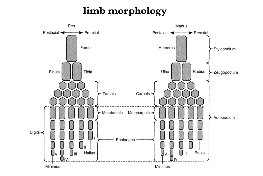
Figure 9.1. Idealized pes (left) and manus (right).
Lissamphibia
Amphibian wrists have three rows of carpals. The first row is the most proximal and comprises the ulnare, intermedium, and radiale (from lateral to medial). The second row consists of centralia, and the distal, third row consists of the carpalia. Four metacarpals are present in the manus, and the fifth digit (the most lateral) has been lost. The ankles are similar to the wrists in having three rows, with the proximal tarsals called the fibulare, intermedium, and tibiale (from lateral to medial). The second row is composed of centralia, and the third row of tarsalia. There are five metatarsals in the foot. Although these general characteristics are common in amphibians, they do not capture all the variation in all groups.
Caudata
Salamanders commonly have phalangeal formulas of 1-2-3-2 or 2-2-3-3 in the manus, although some salamanders exhibit a greater reduction of the digits. The salamander pes usually has four metatarsals associated with the five digits, with a phalangeal count of 1-2-3-3-2. Some salamanders have lost the lateral digit on the pes (digit V).
Anura
Among anurans, the manus usually has four digits, while the pes has five. The manus tends to have the generalized amphibian features described above. The pes has been highly specialized for saltatorial locomotion. The tarsal bones are modified and consist of two rows. The proximal row has a medial astragalus (which represents the fused tibiale, intermedium, and centrale) and a lateral calcaneum (fibulare). These tend to be very elongate and fused at both ends (and sometimes in the middle). The distal row of tarsals is reduced to two or three cartilaginous or bony pieces. The metatarsals and pedal phalanges are long, and the small bone called the prehallux, or calcar, occurs on the tibial side of the tarsus. The typical phalangeal count for the pes is 2-2-3-4-4.
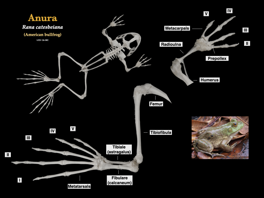
Figure 9.2. Arm and leg details from a bullfrog (Rana catesbeiana, specimen 126-C58 from the UCB teaching collection). Bullfrog photo from Flickr user Will Brown under CC BY 2.0.
Reptilia
Testudines
Turtle hands and feet exhibit substantial differences in the morphology and numbers of the phalanges, which range from the short, stubby digits of tortoises to the long, paddle-like feet of sea turtles.
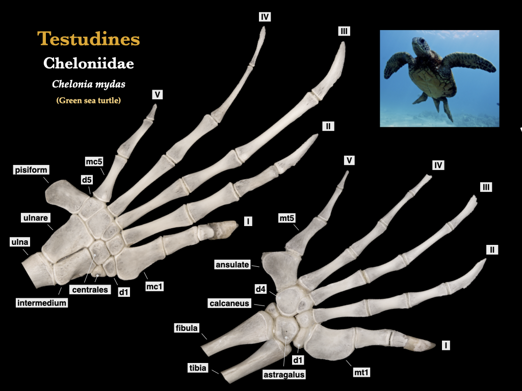
Figure 9.3. Pes (left) and manus (right) from a sea turtle (Chelonia mydas). Note that ansulate is proposed as alternative name for the hooked element in the pes of turtles (Joyce et al. 2013), and that is followed here. The distal tarsal and carpal series is labeled d1–d5. Hildebrand collection specimen (MVZ 890). Green sea turtle photo from Flickr user M Cheng.
Squamata
The pes rays of lizards are often considerably longer than those of the manus, and the phalangeal formulae for pentadactyl squamates are typically 2-3-4-5-3 for the manus and 2-3-4-5-4 for the pes.
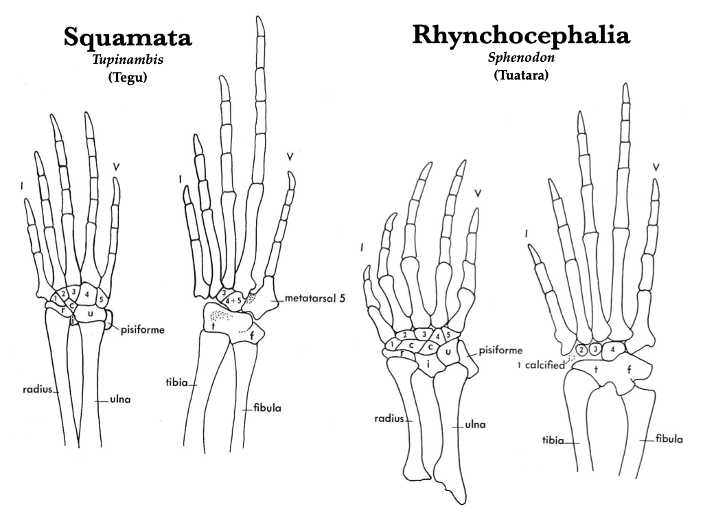
Figure 9.4. Manus and pes of tegu (Tupinambis) and tuatara (Sphenodon) in the following views: right manus of Tupinambis (A), right manus of Sphenodon (B), right pes of Tupinambis (C), right pes of Sphenodon (D). Illustrations from Jollie (1962) under CC0 public domain.

Figure 9.5. Iguana iguana skeleton, with manus (lower right) and pes (lower left). Specimen is from the Hildebrand collection (MVZ 912). Iguana photo from Judy Gallagher under CC BY 2.0.
Crocodylia
In crocodylians, the proximal carpals are elongate and resemble metacarpals with a rotatory joint between the astragalus and calcaneum.
Aves
Birds have four digits on the pes (digits I to V), but only three digits on the manus (digits I to III). The autopodium has been highly modified for flight.
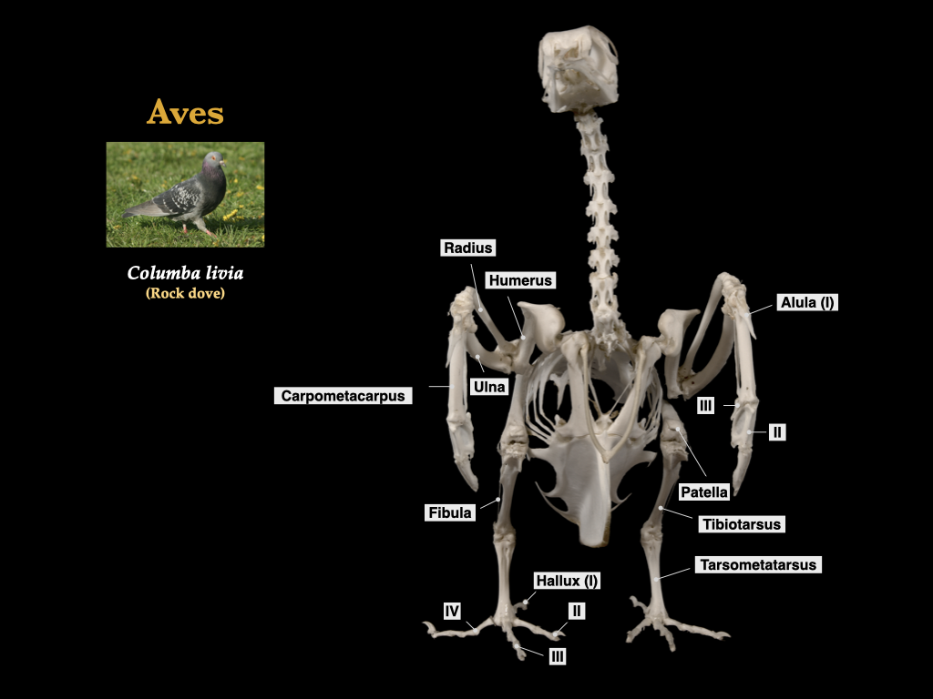 Figure 9.6. Skeleton of a pigeon illustrating the specialized forelimb and hindlimb. Columba livia, UCB teaching collection.
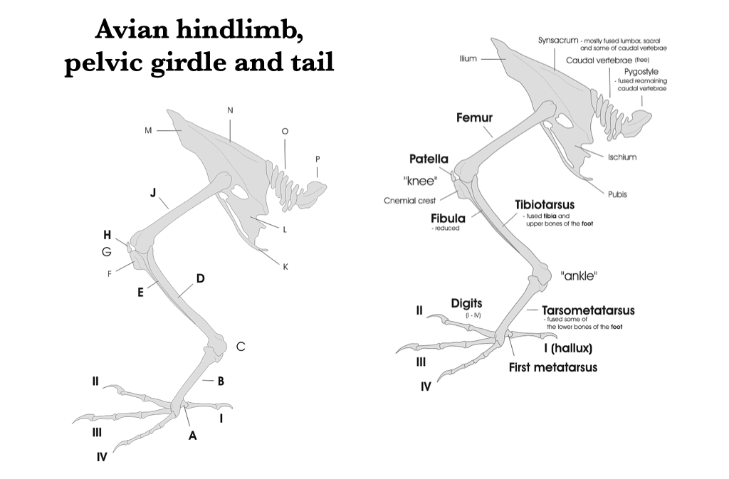
Figure 9.7. Pelvic girdle and hindlimb of a bird from Wikipedia user Darekk2 under CC BY-SA 3.0.
Mammalia
Mammals are specialized in many different ways. Humans, notwithstanding the opposable thumbs and bipedal propensity, do not have radically modified hands and feet.
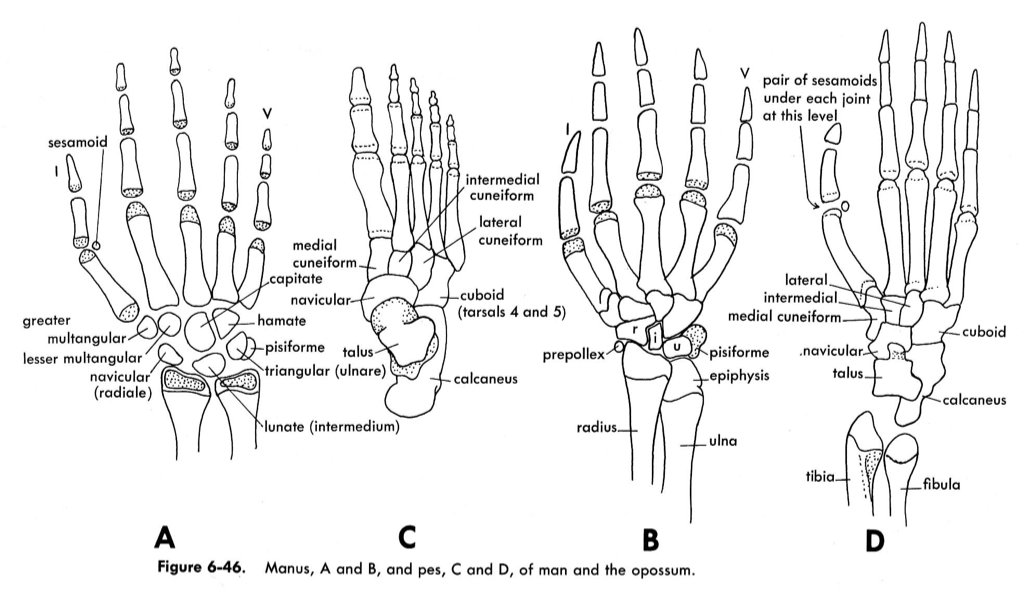
Figure 9.8. Manus and pes of opossum (Didelphis virginiana) and human (Homo sapiens): human manus (A), human pes (C), opossum manus (B), and opossum pes (D). Illustrations from Jollie 1962 under CC0 public domain.
Rodents are diverse in terms of their locomotion, and this is reflected in their autopods. Rodents have seven carpals, but the scaphoid and lunar are often fused, and there is often a radial sesamoid. Fossorial forms tend to have short, broad, robust feet; arboreal climbers, such as squirrels, have a long carpus and tarsus; and ricochetal forms have enlarged hind feet, with the metatarsals fused and reduced in number.
In the carpus of carnivorans, the scaphoid, lunar, and centrale are fused into a scapholunar, and this bone is a synapomorphy of the clade Carnivora. A large pisiform and a small radial sesamoid are present. The first metacarpal is reduced in the Canidae and lacking entirely in Hyaenidae. In seals and sea lions (which have flippers), the first digit is the longest. In general, carnivoran phalanges are relatively waisted (narrow in the middle).
For both carnivorans and ungulates, limb and foot posture are important in shaping manus and pes morphology. There are three main foot and hand postures: plantigrade, digitigrade, and unguligrade. Plantigrade vertebrates walk with the podials, metapodials, and phalanges all on the ground surface (examples include shrews, bears, raccoons, and humans). Digitigrade animals walk with only the phalanges on the ground (examples include cats and dogs). Unguligrade animals walk only on the distal, or ungual, phalanges (examples include most artiodactyl and perissodactyl species).
Artiodactyla includes the ungulates with an even number of toes. Artiodactyls have a unique astragalus characterized by trochleated surfaces that articulate proximally with the tibia and distally with the navicular: the double trochleated astragalus is a synapomorphy of the Artiodactyla. The third and fourth metapodials are the largest, which follows, since the third and fourth digits bear the weight of the body. Most artiodactyls, including deer, antelope, bovids, goats, and giraffes have fused the cuboid and the navicular.
Perissodactyla includes the ungulates with an odd number of toes, such as rhinos, tapirs, and horses. The perissodactyl astragalus has deeply grooved trochlea on the proximal end, and a short, flat distal end. All the weight of horses is borne on the third digits. The carpals and tarsals are flattened and stacked on top of one another, rather than staggered as in artiodactyls.
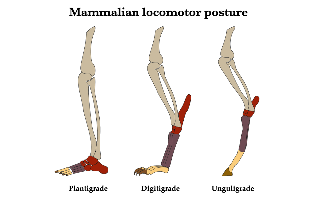
Figure 9.9. Mammalian locomotor posture from Wikipedia user Antoine ADAM under CC0 public domain.
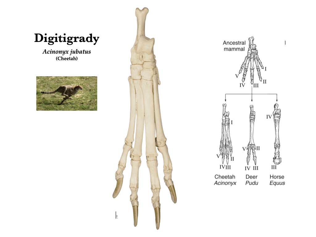
Figure 9.10. Digitigrade pes of a cheetah (Acinonyx jubatus). Cheetah photo from Malene Thyssen under CC BY-SA 3.0; cheetah specimen from the Hildebrand collection (MVZ 1316).
.jpg)
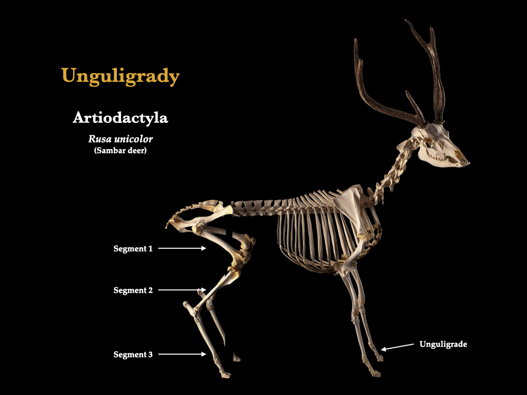
Figure 9.11. Mounted deer skeleton illustrating unguligrade posture. Rusa unicolor specimen from Wikipedia user Museum of Veterinary Anatomy FMVZ USP under CC BY-SA 4.0.
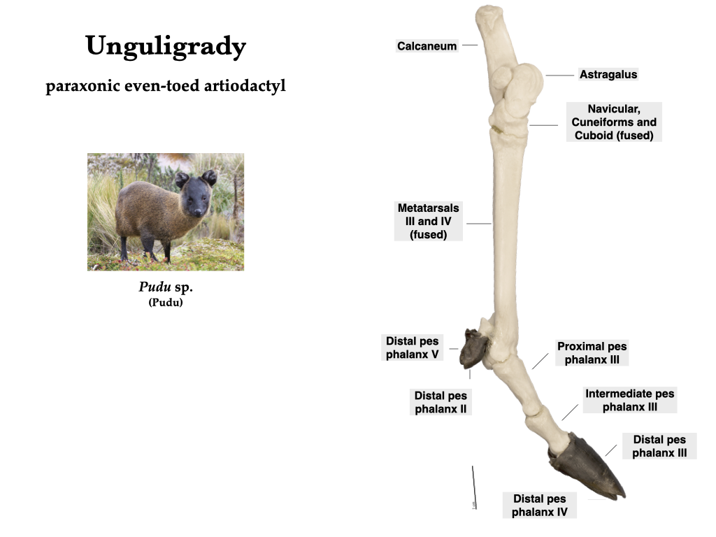
Figure 9.12. Tarsus and pes of an artiodactyl illustrating the unguligrade morphology of an even-toed ungulate. Pudu photo from Flickr user Eider Joselito Chaves Chaves under CC BY 2.0; Pudu specimen from the Hildebrand collection (MVZ 907).
Lipotyphlans usually have seven carpals, but the hedgehog (Erinaceus) has fused the scaphoid and lunar. The lipotyphlan manus is unspecialized, except in moles, where the manus is specialized for digging. Moles have all eight carpals, plus large ulnar and radial sesamoids. Lipotyphlans have unspecialized feet with seven tarsals.
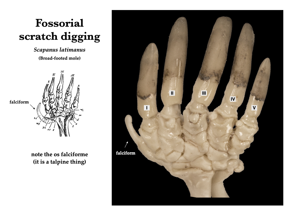
Figure 9.13. Manus of a mole (Scapanus latimanus) illustrating the os falciform (MVZ 840). Illustration on left from Bedard (1902, figure 252) under CC0 public domain.
The hands of chiropterans (bats) are modified into wings. The proximal carpals may be fused to form a single element. The first digit usually does not support the wing membrane, instead it forms a grasping claw. The four remaining digits are wildly attenuated and provide a scaffold for the wing membrane. The feet of bats are less specialized, but they have an elongated calcaneum that attaches to the uropatagium (the membrane in the tail region). A cartilaginous rod called a calcar may extend from the feet to provide additional support to the uropatagium.
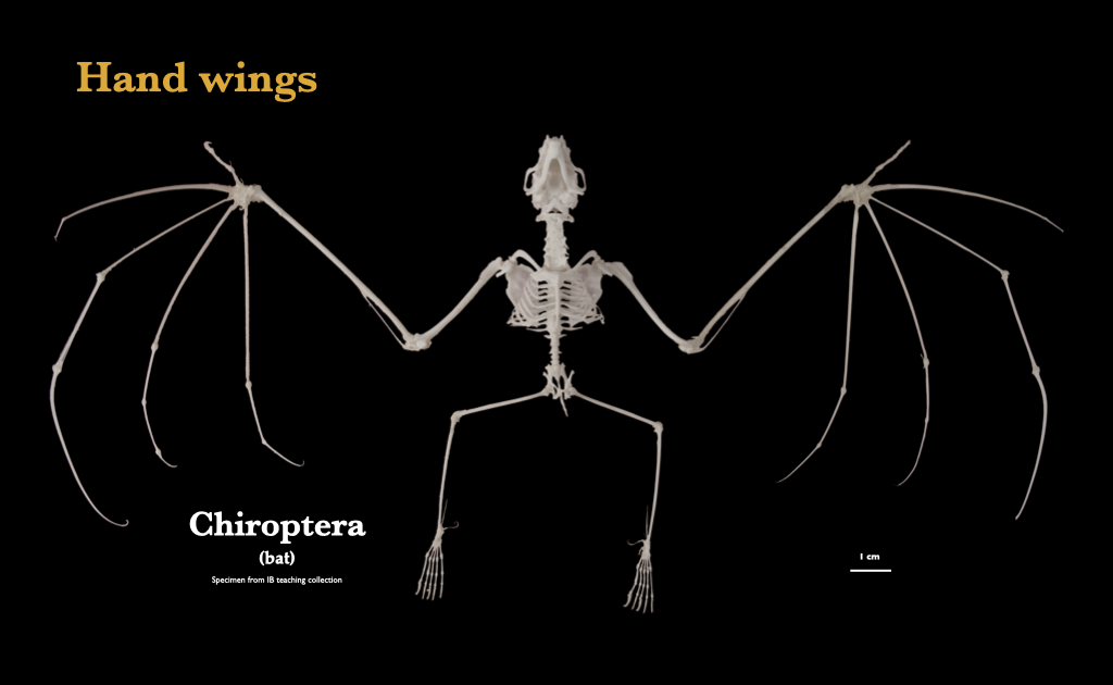
Figure 9.14. Bat wing, showing the extreme reduction of the ulna, and the elongation of the metacarpals and phalanges to support a wing membrane. Chiroptera specimen from IB teaching collection.
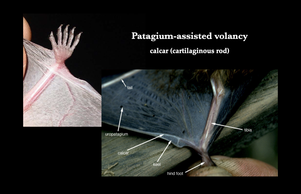
Figure 9.15. The connection of a cartilaginous rod (calcar) to the ankle and uropatagium of a bat. Indiana bat foot image from Wikipedia user USFWSmidwest under CC BY 2.0; Bat calcar image from Wikipedia user Zenofwar under CC BY-SA 3.0.
Metatherians (marsupials) generally have small, shallow astragali. Some marsupials are syndactylous, where digits II and III of the pes are closely appressed, similar in size, and smaller than all the other digits.
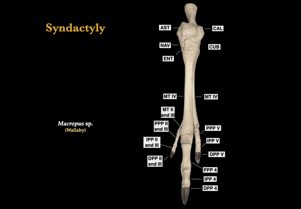
Figure 9.16. Pes of a kangaroo, showing syndactylous toes II and III. Macropus specimen MVZ 769.
The forelimbs of cetaceans are modiffied into flippers, and the number of phalanges may vastly exceed the typical maxima of placental phalangeal counts.
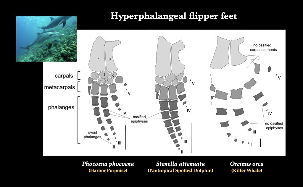
Figure 9.17. Cetacean flippers by Mellor et al. (2009), under U.S. Department of Commerce at DigitalCommons@University of Nebraska – Lincoln.
Primates that rely on swinging by the arms through the tree canopy have radically modified forelimbs for below-branch brachiation. The phalanges are long and curved, and the stylopod and zeugapod are very long and thin overall.
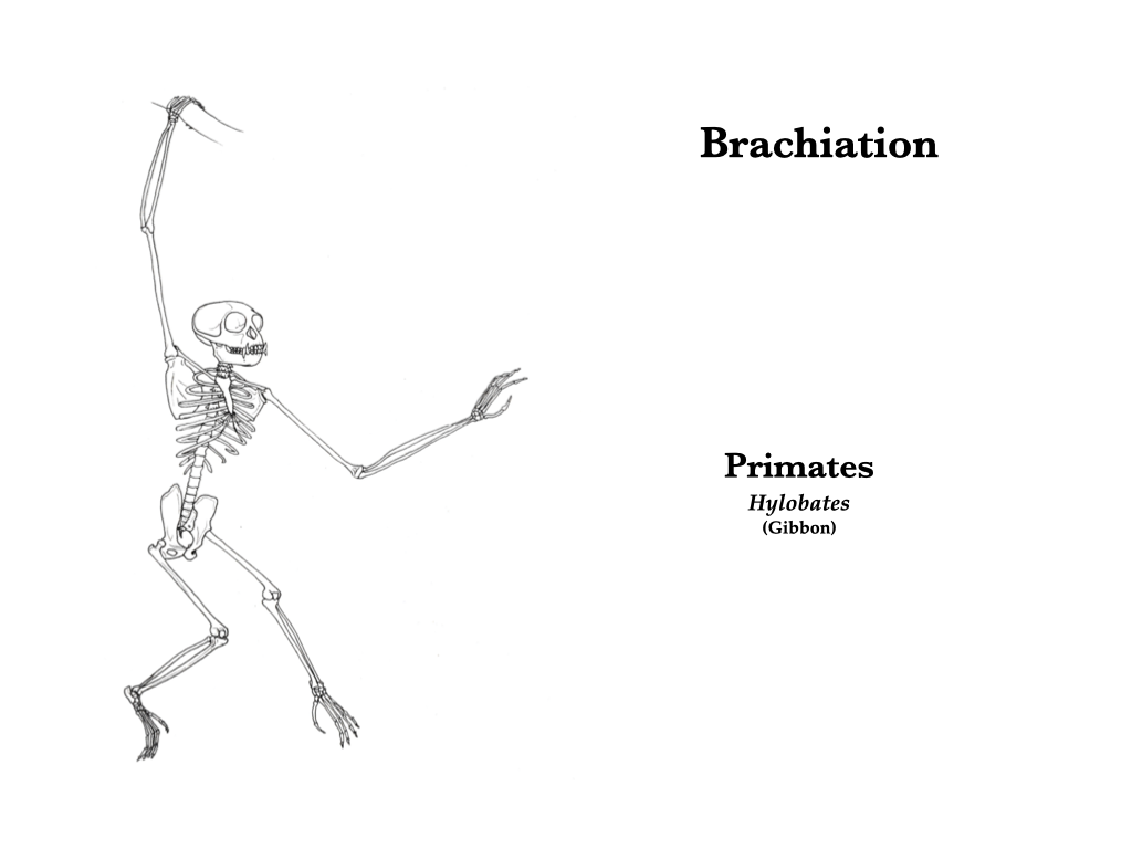
Figure 9.18. Brachiation in a gibbon.