5 Teeth
Objectives
- Learn basic tooth anatomy and terminology.
- Identify the types of teeth in heterodont mammalian dentition and identify the cusps and features of molar teeth (with particular emphasis on tribosphenic molars).
- Describe the dental formula of a dentition and use dental formulae to distinguish marsupials from placentals.
- Identify the specialized tooth types associated with major dietary types.
- Use tooth morphology to predict dietary preference and ecological function.
Overview
In this lab, we will study the evolution of the tetrapod feeding apparatus with a focus on tooth morphology. Teeth vary in structure and attachment to the skeleton depending on the animal’s diet and feeding mechanism. For the majority of vertebrate taxa, teeth are homodont, meaning that they are structurally similar (though they may vary in size). Many fish and non-mammalian tetrapods have simple, conical teeth that attach to the jaws superficially via connective tissue—the acrodont and pleurodont conditions. Acrodont and pleurodont teeth are effective in capturing and handling small prey without mastication. In the evolution of the tetrapod feeding apparatus, the teeth became specialized for particular functions, and teeth took on divergent roles based on their position in the dental arcade (heterodonty). The heterodont teeth of mammals became more solidly attached to the skull by growing firmly in a socket (thecodonty). In this lab, you will learn how to use vertebrate tooth morphology to interpret ecological function and to classify specimens based on functionality. You are responsible for terms listed in bold face in the text and terms lists. For your reference, Terms List 5.2 provides examples of taxonomic groups that display the common trophic types.
See the terms list here: Terms 5.1–5.2
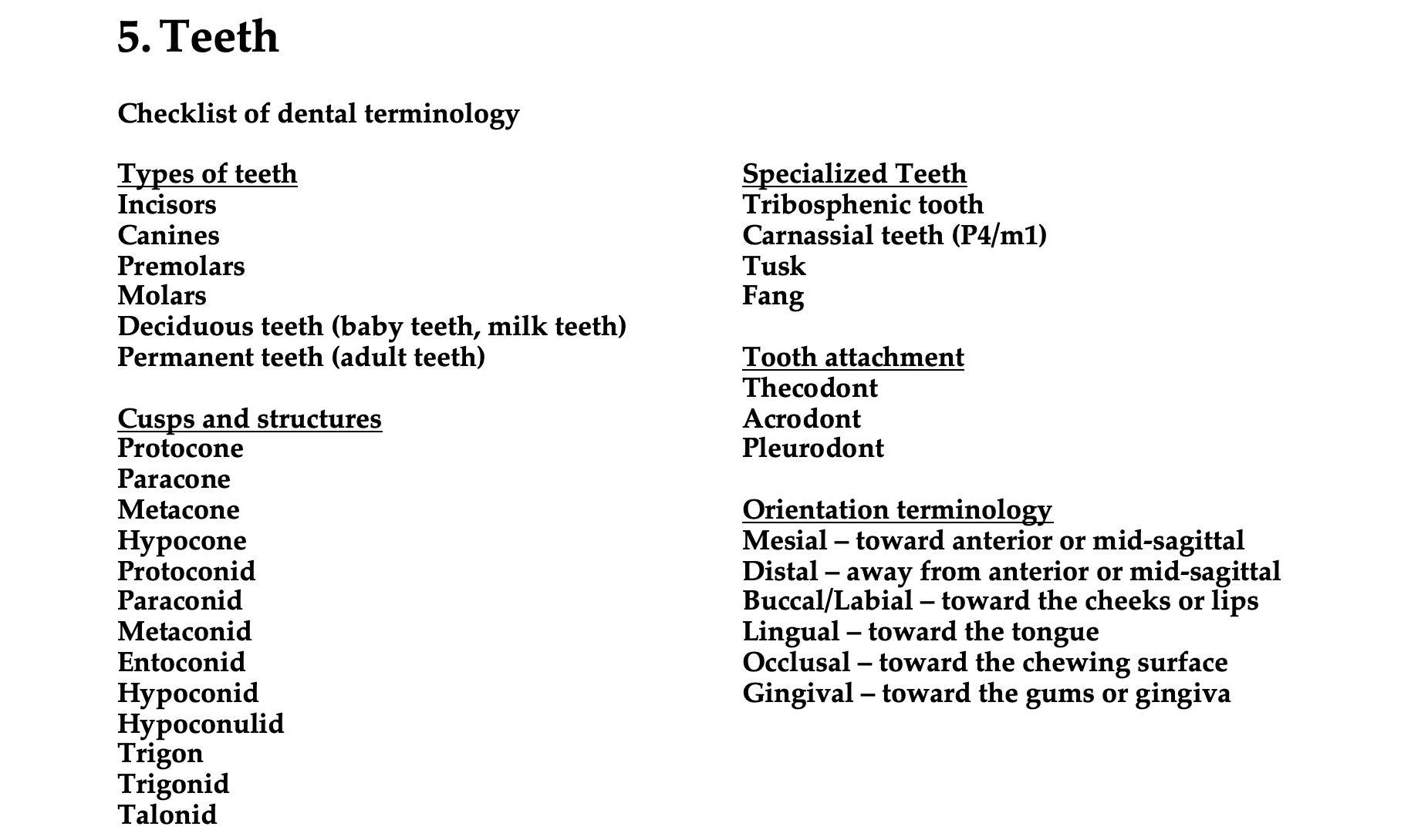
Orientation terminology
Mesial – toward anterior or mid-sagittal
Distal – away from anterior or mid-sagittal
Buccal/Labial – toward the cheeks or lips
Lingual – toward the tongue
Occlusal – toward the chewing surface
Gingival – toward the gums or gingiva
Directional terminology in the oral cavity
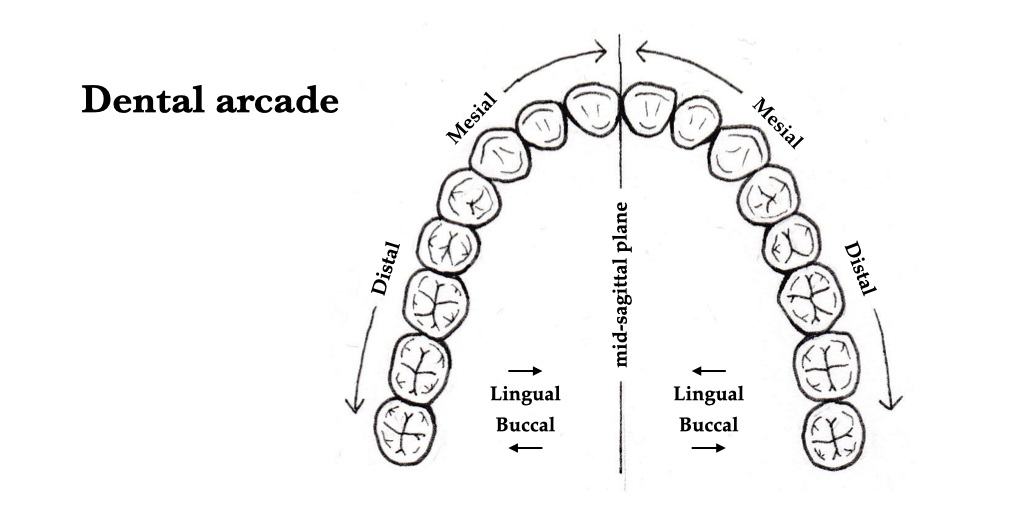
Figure 5.1. Directional terms to describe relative position in the oral cavity.
Additional terminology of the dentition
Most non-mammalian vertebrates have homodont dentition, in which all of an individual’s teeth are structurally and morphologically similar. Some mammals, such as toothed whales (odontocetes) and armadillos (Dasypus) also have homodont dentition, but these species generally do not chew their food. Other tetrapods, such as termite-eating mammals (myrmecophages), baleen whales, and turtles are edentulous and lack teeth altogether. Most mammals, however, have heterodont dentitions, in which there are three or four different types of teeth that are specialized to perform distinct food processing functions.
Heterodont – Having different parts of the dentition morphologically specialized to do different things. Most mammals are heterodont.
Homodont – Having an undifferentiated dentition, although tooth size usually varies across the row. Sharks, crocs, and toothed whales are homodont.
Edentulous – Lacking teeth. Turtles, anteaters, and baleen whales are edentulous.
Diastema – A naturally occurring gap between teeth of different function, usually used with reference to the gap between the anterior teeth and the cheek teeth. All rodents and horses exhibit a diastema.
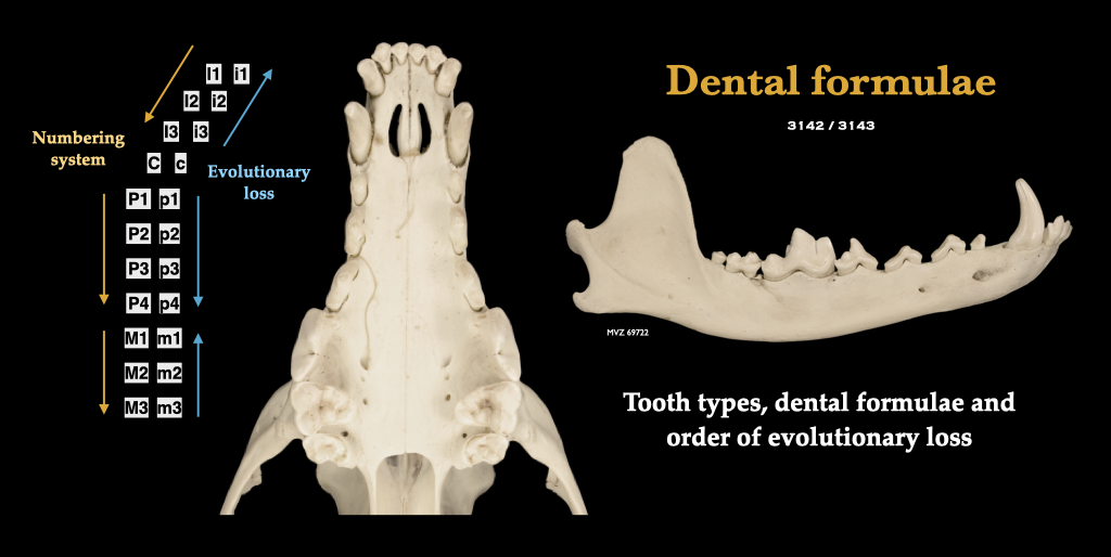 Figure 5.2. The dentition of a coyote to illustrate the types of teeth, the dental notation system, and the pattern of tooth loss over evolutionary time. Canis latrans, MVZ 69722.
Dental formula
Mammalian vertebrates are marked by a dentition that is heterodont, with distinct incisors, canines, premolars, and molars. Note that the teeth are numbered from front to back (mesial to distal), but that teeth are lost in evolution from back to front (in the incisors and molars) or from front to back (in the premolars). A dental formula is a convenience for expressing the number of each tooth type in each quadrant of the upper and lower jaws of mammals. It is expressed as a string of numbers (representing one quadrant of the upper dentition), separated by a slash, followed by another string of numbers (representing one quadrant of the lower dentition). For example, the dental formula of most dogs is 3142/3143. Dental formulae vary systematically across mammals in relation to diet and phylogeny.
Marsupials and placentals differ fundamentally at the level of the dental formula. The maximum number of teeth in the mouth of a marsupial mammal is 50, with a formula of 5134/4134, illustrating the maximum number of teeth of each type (note that there may be up to five upper incisors in marsupials). The maximum number of teeth in the mouth of a heterodont placental is 44, with a formula of 3143/3143 (note that there can be no more than three incisors in placentals). These maximal formulae represent the primitive conditions, and evolution is marked by reductions from these maxima over time. Note that the common opossum (Didelphis virginiana) exhibits the primitive dental formula for the marsupials, and moles (Scapanus latimanus) and pigs (Sus scrofa) exhibit the primitive formulae for the placentals.
In the heterodont dentition of a mammal, incisors are the most mesial teeth, and they are typically small and spade shaped. Incisors are effective in cutting and securing food, as in horses, or for gnawing, as in rodents. The upper incisors are always rooted in the premaxillae. Canines are distal to the incisors and they are basically conical in shape. Canines are adapted for seizing and piercing prey, and for fighting. The upper canines are rooted either on the suture between the premaxilla and maxilla, or just distal to the suture. The teeth distal to the canines are the premolars, and the molars are distal to the premolars. Premolars and molars are differentiated by their ontogenetic replacement pattern. Premolars appear in both the deciduous and permanent sets of teeth, whereas molars occur as permanent teeth and do not have deciduous precursors (molars are monophyodont).
Ancestrally, the premolars were for piercing and the molars served a dual purpose of cutting and crushing, but a lot of variability and complexity has evolved in these teeth. Often, the premolars are morphologically similar to the molars, in which case they are collectively referred to as “cheek teeth.” Most cheek teeth have complex patterns of cusps and ridges to aid in the reduction of difficult-to-process plant matter. The molars of the earliest mammals had three cusps that were in line with one another from mesial to distal. In more derived early mammals, the cusps shifted so that there was a single, large cusp (apex) on one side of the tooth and two small cusps on the other. In the upper teeth, the apex was shifted lingually (trigon configuration) whereas on the lower molars, the apex was shifted buccally (trigonid configuration), such that the apex of the upper molar sheared against the paired cusps on the lower molar and vice versa. The protocone evolved as an additional cone on the lingual side of the upper molar in the ancestor of marsupials and placentals. Of the two cusps on the buccal side of the upper molar, the mesial one is the paracone and the distal one is the metacone. On the lower molar, the apex is called the protoconid, and the more mesial of the paired cones on the lingual side is the paraconid, and the distal cone is the metaconid. These three cusps together make up the trigonid. Distal to the trigonid is a low, shallow basin, edged by three distal cusps, from lingual to buccal: entoconid, hypoconulid, and hypoconid. This low platform behind the trigonid is called the talonid. Molars of this teeth are called tribosphenic, and during occlusion, the upper molar shears against the trigonid, and the protocone lands in the talonid basin, creating cutting and crushing actions. Molars of this type are called tribosphenic, and they have been referred to by engineers as “teeth of perfection” given their efficiency in mastication, particularly the mastication of chitinous insect exoskeletons.
Generations of teeth
Definition: An animal that has only a single set of teeth throughout life is referred to as monophyodont. Monophyodonty is the condition observed in odontocetes (e.g., porpoises and dolphins) and in some marsupials. Animals with teeth that are continually developed and replaced throughout life are polyphyodont. Most tetrapods, including crocodilians, exhibit polyphyodonty. Animals that have two successive dentitions during life—a deciduous/milk/baby set and an adult/permanent set—are diphyodont. Most placental mammals, including humans, exhibit diphyodonty.
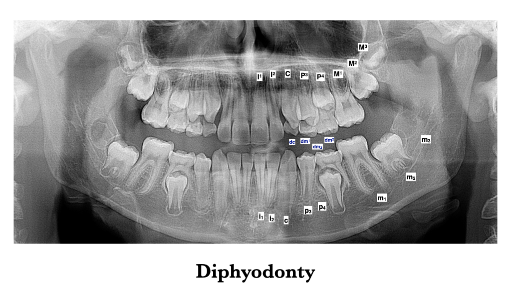
Figure 5.3. Diphyodonty as illustrated in the mouth of an 11-year-old human male.
Tooth anatomy
Teeth are by far the hardest part of the skeletal system, making them more abundant than any other skeletal element and extremely useful for paleontology. Structurally, the tooth has two parts: the crown, which projects above the gum line (gingiva), and the base, which does not. If the base of the tooth is not embedded in a bony socket, the dentition is described as acrodont. If the base of the tooth is embedded in a bony socket (alveolus), it is instead called a “root,” and the dentition is described as thecodont, which is the condition of mammals. The surface of the tooth crown is coated by a layer of hard, crystalline enamel. The surface of the root is composed of a cementum which helps to weld the tooth in the bony jaw. Deep to the enamel and cementum is dentin (or dentine), which is of intermediate hardness relative to enamel (hardest) and bone (softest). Enamel, cementum, and dentin surround a pulp cavity that houses nerves and blood vessels. The occlusal surface of a tooth is the part of the crown that occludes (makes physical contact) with the teeth in the opposing jaw. On the occlusal surfaces are cusps—raised mounds, peaks, and prominences—the morphology of which is critical in tooth function.
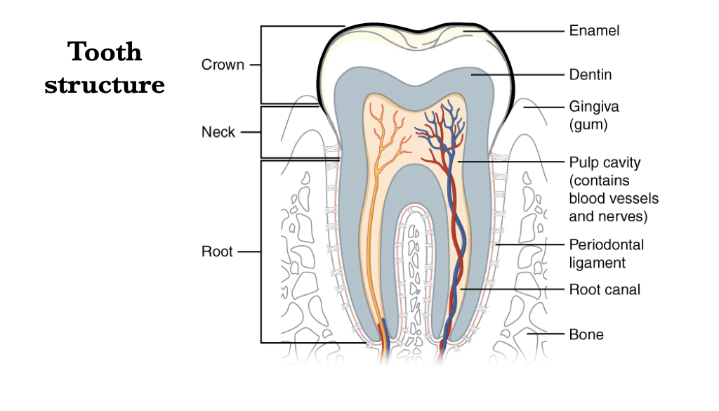 Figure 5.4. Internal structure of a human molar. Image from Anatomy & Physiology, Connexions Web site and OpenStax College under CC BY 3.0.
Tooth attachment to the jaws
Acrodont teeth attach to the jaw with or without a small socket on the crest of the bone; acrodonty is observed in snakes and some lizards. Pleurodont teeth attach to the jaw on the medial surface of the bone, sometimes with a small socket, but usually without; many lizards have pleurodont teeth. Finally, thecodont teeth are set embedded in a socket (alveolus); all mammalian teeth are thecodont.
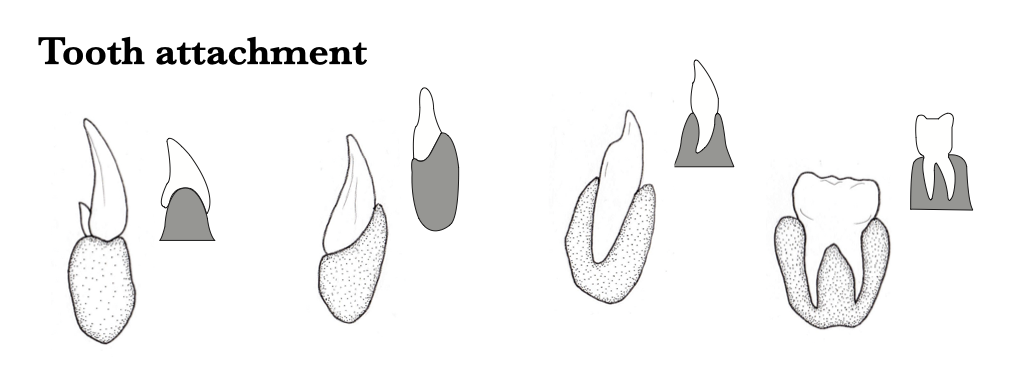 Figure 5.5. Illustration of tooth-to-jaw attachment types, from left to right: acrodont, pleurodont, thecodont with a single root, and thecodont with more multiple roots.
Tribosphenic teeth
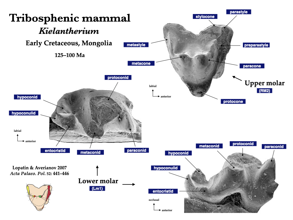
Figure 5.6. Upper and lower tribosphenic teeth of a Mesozoic mammal, Kielantherium. Mesozoic tribosphene from Lopatin and Averianov (2007) under fair use guidelines.
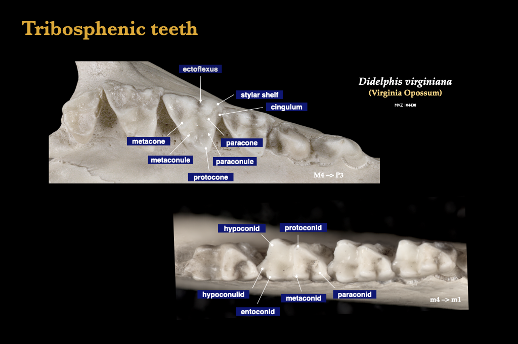
Figure 5.7. Tribosphenic molars of the common opossum (Didelphis virginiana, MVZ 104438).
Cusp morphology
The tribosphenic molars is considered to be the primitive form from which all modern mammalian molars evolved. The cusps of the tribosphenic molars include the characteristic three of the upper trigon (protocone, paracone, metacone), the three of the lower trigonid (protoconid, paraconid, metaconid), and the three of the talonid (entoconid, hypoconulid, and hypoconid). The tribosphenic pattern is understood to be the cusp arrangement from which modern mammals descended, and cuspal homologies have been described across taxa. Try to identify individual cusps during your study of the major occlusal patterns that mark the mammals. As always, connect the morphology of the structures (the form of the teeth) with their ecological role (dietary function).
Tribosphenic teeth are extremely effective in the reduction of insect chitin, and insectivory is thought to be the dietary habit of many of the early modern mammals of the Mesozoic Era. Many extant mammalian insectivores exhibit a modification of this pattern, such that a shearing crest develops in the form of a buccal ectoloph. In the dilambdodont configuration, the cusps for a W-shaped ectoloph, and this is the commonly observed in shrews (e.g., Sorex), moles (e.g., Scapanus), and many bats (e.g., Myotis).
Another modification of the tribosphenic molar pattern occurs with the addition of a fourth major cusp on the upper molars, the hypocone. A four-cusped molar such as this is referred to as quadritubercular, and the hypocone has evolved independently in many mammal lineages. Quadritubecular teeth with cusps that are low and rounded, and good for crushing and grinding, are called bunodont. Mammals with bunodont teeth include raccoons (Procyon), pigs (Sus), bears (Ursus), and many primates, including humans (Homo). The common dietary proclivity of bunodont mammals is omnivory.
Mammals that consume large amounts of vertebrate flesh are called, of course, carnivores. The teeth of carnivores tend to have a significant shearing component, and in the members of the clade Carnivora, this shearing surface is best developed on the upper fourth premolar and the lower first molar, the so-called carnassial pair (P4/m1). Teeth such as these that are designed for shearing meat are called secodont, and they are seen in the carnivorous dogs (Canidae) but they are even more developed in the hypercarnivorous cats (Felidae).
Some of the most complex cuspal patterns are observed in the herbivorous mammals. In selenodont herbivores, the individual cusps are elongated mesiodistally into crescent-shaped crests called selenes. The folds between the selenes run labiolingually, and the jaw is swung from side to side (mediolaterally). Selenodont teeth are seen in deer (Cervidae), cows and antelope (Bovidae), and camels (Camelidae). In other herbivorous mammals, the individual cusps are connected labiolingually, and sometimes mesiodistally, to form a series of ridges called cross-lophs or, simply, lophs. The jaws of lophodont mammals often operate in an anteroposterior direction (perpendicular to the axis of the lophs). Rhinoceroses (Rhinoceratidae) are lophodont, as are many rodents (e.g., Rattus), and both horses and elephants exhibit types of lophodonty.
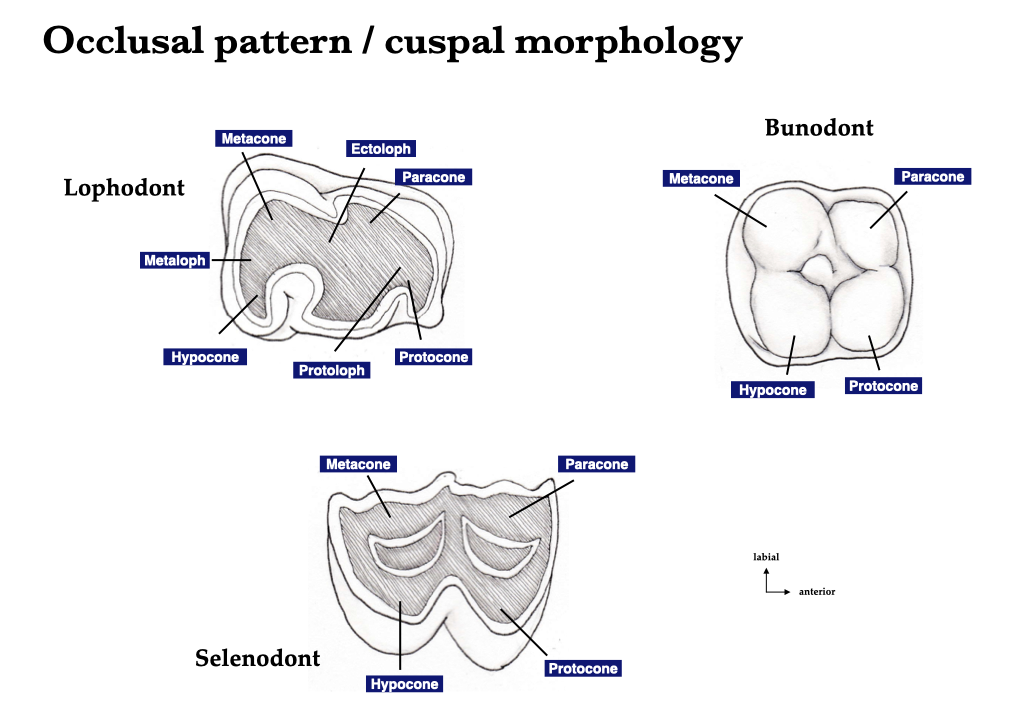
Figure 5.8. Occlusal anatomy and cuspal structure of herbivores (lophodont, selenodont) and omnivores (bunodont).
Tooth crown height
Where the roots of the tooth end, the crown begins (or where the crown ends, the roots begin). The junction between the roots and the crown is called the neck or cervix, and this may occur at gum line or within the dental alveolus. Teeth that have relatively low crowns—where crown height is less than crown length or crown width—are called brachydont. Most bunodont teeth are also brachydont, and this is true for human teeth (Homo sapiens). Teeth that are high-crowned, where crown height is greater than crown length or crown width, are called hypsodont. Many herbivores have hypsodont teeth, including cows and many rodents. A specific case of hypsodonty exists in which the teeth grow continuously throughout life, as opposed to the finite growth process that is observed in brachydont species. Hypsodont teeth that grow continuously in this way are called hypselodont (or, simply, elodont). The cheek teeth of horses grow throughout life and are considered hypselodont, as are the gnawing incisors of rodents.
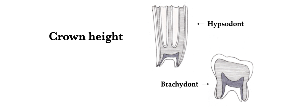
Figure 5.9. Crown height relative to crown length and crown width: high-crowned (hypsodont) and low-crowned (brachydont) teeth.
Specialized tooth types
Tusks are also a modified kind of tooth. Elephant tusks are elongate incisors, whereas walrus and boar tusks are canines. What type of tooth is the tusk of a narwhal?
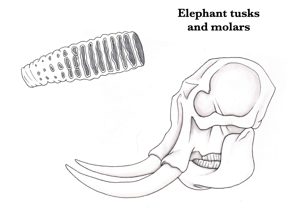 Figure 5.10. Molar tooth (upper left) and incisal tusks (lower right) of an elephant.
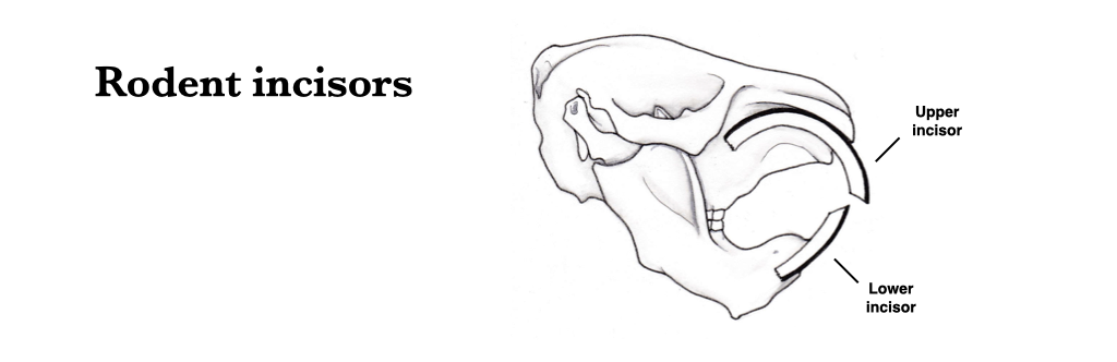
Figure 5.11. The gnawing incisors of rodents form deep within the cranium and mandible and continuously grow as they wear at the tips.
Fangs are another type of modified tooth that are specialized for venom delivery in some snakes. Fangs are located on the maxillae and are defined by being hollow so that venom can pass through them to be injected directly into the prey.
Ecomorphology of teeth
Insectivory
Insect bodies are strengthened by chitin, and vertebrates that evolved to eat insects have teeth that are specialized to crunch, mash, and shear the insect exoskeletons. The tribosphenic tooth is a great example of this (described above). Dilambdodont teeth are marked by a W-shaped ectoloph ridge as seen in the molariform teeth of shrews, talpid moles, and many bats.
Carnivory
In carnivorans, the P4 and m1 contact one another during mastication, and they are referred to as the carnassials. Most carnassial pairs are effective at slicing flesh, and they maintain high shearing surfaces (secondonty). In secodont carnassials, the protocone of the P4 is typically reduced, and the metaconid and talonid of the m1 are reduced. Cats are hypercarnivores (heavily reliant on meat), and they elongate the shearing component of their carnassials by adding a parastyle to the P4, and they lose the talonid on the lower first molar completely, giving them a distinctive butterfly-shaped m1. Dogs are less specialized for a strict meat diet and they rely on a well-developed talonid on the m1 for crushing and grinding food.
Durophagy
Durophagy refers to the consumption of hard-shelled organisms. Durophagous vertebrates rely on heavy jaws and specialized teeth to crush their prey orally. The molariform teeth are stout with broad, short crowns, thick enamel, and well-developed roots set in dense bone. Powerful temporalis muscles help to generate massive crushing force. Two great examples of durophagy available for study in the lab are the water tegu (Dracena) and the sea otter (Enhydra lutris).
Osteophagy
Osteophagy is the habit of eating bones. Bones are very hard objects, and osteophages, like durophages, require specialized crushing jaws and teeth. The best modern examples of osteophagy are the bone-crushing hyenas (Crocuta, Hyena). In these hyenas, the p3 and P2/P3 work together to smash heavy bones up to the size of giraffe tibiae. The osteophagous hyenas maintain a second carnassial pair for the efficient processing of flesh as well.
Herbivory
Herbivory is defined as the consumption of vegetation, and plant matter is often fibrous, rich in phytoliths and silica, or otherwise abrasive and difficult to reduce. In other words, the herbivorous diet can create heavy wear on occlusal surfaces. Many herbivores have teeth with tall crowns (hypsodont) or tooth crowns that grow continuously, replacing worn tissue when worn (hypselodont). Herbivore tooth cusps tend to be connected with ridges to form selenes (ridges that are mesiodistally oriented) or lophs (ridges that are labiolingually oriented). Occlusal ridges and crests are generally oriented perpendicular to the dominant chewing motion of the jaw. This creates a surface on which plant material can be heavily ground. Herbivore premolars tend to become large and molariform, which adds to the overall grinding area of the cheek teeth.
Omnivory
Omnivores prepare food by pounding, rather than by shearing. Typically, a hypocone is added to the upper cheek teeth and the paraconid is lost from the lower cheek teeth. Bunodont dentition is typical of omnivores, as seen in bears (Ursidae), some pigs (Suidae), raccoons (Procyonidae), and primates.